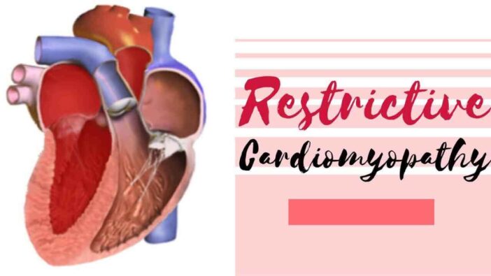
What is Restrictive Cardiomyopathy?
Restrictive cardiomyopathy is a rare type of cardiomyopathy (a problem of the heart muscle). More clearly, this condition describes the heart ventricles that are stiff and less flexible to expand when filled with blood.
People with this condition have a heart that can’t pump blood properly. Blood from the heart cannot get out of the rest of the body. As a result, the ventricles and atria will enlarge and can cause heart failure.
In some cases, this condition also makes fluid accumulate in the body, including the lungs. Heart disease that attacks the ventricles has many other designations, namely infiltrative cardiomyopathy or idiopathic restrictive cardiomyopathy.
Restrictive Cardiomyopathy Causes
In many cases, the cause of RCM is unknown. However, RCM can be caused by genetic factors and run in families. In 30% of patients, there is a family history of RCM disease. Another cause is the buildup of scar tissue, abnormal proteins, or iron in the heart muscle. Certain cancer treatments, such as radiation therapy and chemotherapy can also cause RCM. Sarcoidosis, a disease that causes inflammation of body tissues is also the cause of RCM.
Restrictive Cardiomyopathy Symptoms
RCM often causes no symptoms before it worsens. The patient will begin to feel fatigue and shortness of breath. Other symptoms are fainting and dizziness. A cough that does not go away and an abnormal heartbeat are also very common.
In pediatric patients, the first symptoms of RCM are usually related to lung problems. Asthma or chronic lung infection are symptoms that often appear in pediatric patients. Other symptoms are fluid buildup in the hands, feet, face, and abdomen, as well as swelling of the liver. In such cases, most patients get treatment from a respiratory specialist or gastroenterologist first. Then, they were referred to a heart specialist because X-rays showed heart abnormalities.
Restrictive Cardiomyopathy Complications
Complications of restrictive cardiomyopathy include [1,2]:
- Thromboembolism.
- Stroke due to blood clots in the brain.
- Dysrhythmia.
- Heart failure.
- Cirrhosis of the heart.
- Extra manifestations of cardiac, depending on the main cause.
- Increased risk of pregnancy complications
- Sudden cardiac death due to a dangerous heart rhythm.
Restrictive Cardiomyopathy Diagnosis
The size of the heart can remain normal in restrictive cardiomyopathy conditions. In some cases, restrictive cardiomyopathy can be difficult to distinguish from constrictive pericarditis, a condition in which the pericardium layer (liver wrapping membrane) undergoes thickening, calcification, and stiffness. [3]
The condition of constrictive pericarditis prevents the heart muscle from enlarging during filling time and affects heart function. Certain diagnostic tests need to be done by people with restrictive cardiomyopathy to eliminate the diagnosis of pericarditis and confirm the diagnosis of restrictive cardiomyopathy. [3]
Some other differently identified diagnoses in cases of restrictive cardiomyopathy are[1]:
- Acute or chronic heart failure
- Hypertensive heart disorders
- Hypertrophic cardiomyopathy
- Acute or chronic pericarditis
Restrictive cardiomyopathy can be diagnosed based on personal medical history, family medical history, physical examination and diagnostic tests.
When performing a physical examination, the doctor may notice extra cardiac manifestations, such as carpal tunnel syndrome, which can appear in people with amyloidosis or sarcoidosis. Some of the possible tests are [1,2,3,4]:
- Blood tests, to help determine the type of restrictive cardiomyopathy.
- Electrocardiogram, to check the rhythm of the heart.
- Chest X-ray, to see the anatomy and size of the heart.
- Echocardiogram, to check blood flow in the heart and find out the heart’s ability to pump blood throughout the body.
- Treadmill test (exercise stress test), to find out the condition of the heart when exercising.
- Cardiac catheterization with coronary angiography, to see the condition of the arteries and measure pressure in the heart.
- CT scans, to see the condition in the body.
- MRI, to look at the anatomy of the heart and coronary arteries.
- Radionuclide studies (nuclear imaging), to look at affinity with people with amyloidosis.
- Biomarkers, such as troponin T, BNP (B-type Natriuretic peptide), and pro-BNP, to assist in diagnostics and determining prognostic factors
A myocardial biopsy may also be performed to check for the cause of restrictive cardiomyopathy. During a myocardial biopsy, small tissue is taken from the heart and examined under a microscope to determine the cause of the symptoms. [3]
Medical Research & Source
- Kristen N. Brown, Venkata Satish Pendela, Rene D. Diaz. Restrictive Cardiomyopathy. StatPearls; 2021.
- Steven Kang, MD & Lu Cunningham. Restrictive Cardiomyopathy. Cedars Sinai; 2021.
- Cleveland Clinic medical professional. Restrictive Cardiomyopathy. Cleveland Clinic; 2019.
- Steven Kang, MD & Lu Cunningham. Restrictive Cardiomyopathy. Cedars Sinai; 2021.
- Image: Npatchett, CC BY-SA 4.0 https://creativecommons.org/licenses/by-sa/4.0, via Wikimedia Commons
- Video: Bryte Medical, M.D, CRNA, RGN
![Liver Abscess: Causes, Symptoms and Treatment [Full Explanation] 1 Liver Abscess: Causes, Symptoms and Treatment [Full Explanation]](https://markethealthbeauty.com/wp-content/uploads/2022/07/Liver-Abscess-300x168.jpg)

