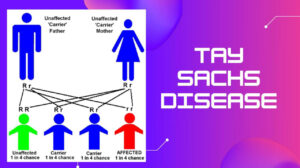
Markethealthbeauty.com – Spinal muscular atrophy (SMA) is a group of congenital abnormalities that cause progressive muscle degeneration. SMA is the second leading cause of neuromuscular diseases inherited due to the recessive nature of autosomal damage from both parents. The SMA is estimated to affect only 1 in 6,000- 10,000 people.
What Is Spinal Muscular Atrophy?
Spinal Muscular Atrophy (SMA) is a neuromuscular genetic disorder characterized by muscle paralysis. The real clinical appearance of SMA patients is muscle paralysis, especially in both legs. However, the main source of paralysis is not caused by damage to muscle cells, but rather pure paralysis caused by damage to nerve cells in the spinal cord.
The spinal cord is a part of the central nervous system that runs continuously from the brain down to the lower back.
From this spinal cord, the branches of innervation are responsible for every part of the body, including the limbs and feet.
As we know, muscle movements are controlled by the brain by the intermediation of the spinal cord, where the nerves connecting the brain with the muscles pass through the spinal cord.
Thus, it can be understood that damage to nerve cells in the spinal cord leads to loss of motor control capabilities, especially in the muscles responsible for movements such as crawling, walking, chewing, head and neck control, even breathing.
In this case, the leg muscles and breathing are more frequent and more severe paralyzed than other muscles.
Paralysis causes muscles to never be used, thus making it shrink (atrophy), especially clearly visible on the legs.
Spinal Muscular Atrophy Types
Based on its severity, SMA is divided into three types.
SMA Type I
SMA Type I, also known as Werdnig-Hoffmann Disease, is the most severe type.
Symptoms in Type I high school begins very early, either before birth or at the latest from the age of 6 months after birth. The symptoms are characterized by difficulty breathing, inability and complete muscle weakness.
The main problem in Type I SMA babies is weakness in the respiratory muscles, which makes them often depends on respiratory aids. Babies with SMA Type I have a very low life expectancy, where all or almost all die before the age of 2 due to respiratory failure.
SMA Type II
SMA Type II has less severity, when compared to type I.
SMA symptoms in type II start between the ages of 6 to 18 months.
Children with type II SMA can sit unaided and can sometimes stand painstakingly holding on to their feet. But none of them can walk.
Although his life expectancy is higher than that of type I SMA, in general children with type II SMA experience severe respiratory problems that are the cause of death in early childhood.
SMA Type III
SMA type III, also called Kugelberg-Welander Disease, is the type with the lowest severity.
The symptoms only begin at the age after 18 months.
It usually begins with normal motor development and then in early childhood experiences a significant decrease in motor ability. In rare cases, new symptoms begin to appear in adulthood (some experts call it SMA Type IV).
Spinal Muscular Atrophy Symptoms
The symptoms of SMA and when it first appears to depend on the type of SMA you have.
Typical symptoms include:
- Drooping or weak arms and legs
- Movement problems – such as difficulty sitting, crawling or walking
- Muscle twitching or shaking (tremor)
- Bone and joint problems – such as unusually curved spine (scoliosis)
- Swallowing problems
- Difficulty breathing
SMA does not affect intelligence or the cause of studying disability.
Spinal Muscular Atrophy Treatment
It is currently impossible to cure SMA, but research is ongoing to discover the possibility of new treatments.
Care and support are available to manage symptoms and help people with SMA have the best quality of life.
Treatment may involve:
- Exercises and equipment to aid movement and breathing
- Tube food and diet advice
- Braces or surgery to treat problems with the spine or joints
A variety of health care professionals may be involved in your treatment, including specialists, physiotherapists, occupational therapists, and speech and language therapists.
Source:
- Image: Cervical vertebra blank.svg: Fred the OysterPolio spinal diagram.PNG: DO11.10Derived: Angelito7, CC BY-SA 4.0 https://creativecommons.org/licenses/by-sa/4.0, via Wikimedia Commons.
- Video: ASGCT


